


A look at the selected microscopic time-lapses in Nikon’s “Small World, in Motion” competition
The selected works of the “Nikon Small World” competitions were introduced in the last months of 2019 in the two sections of photomicrography and microscopic time-lapses.
To watch the selected works in the microscopic time-lapse section of this competition.
“Small World in Motion” is the title of a part of “Nikon’s Small World” competition, which has hosted the works of artistic scientists every year since 2011. In this section, recorded time-lapses compete with optical microscopes. To watch these competitions’ selected works of 2019, stay with Zomit in the two areas “Selected Videos” and “Special Appreciation.”
Selected videos
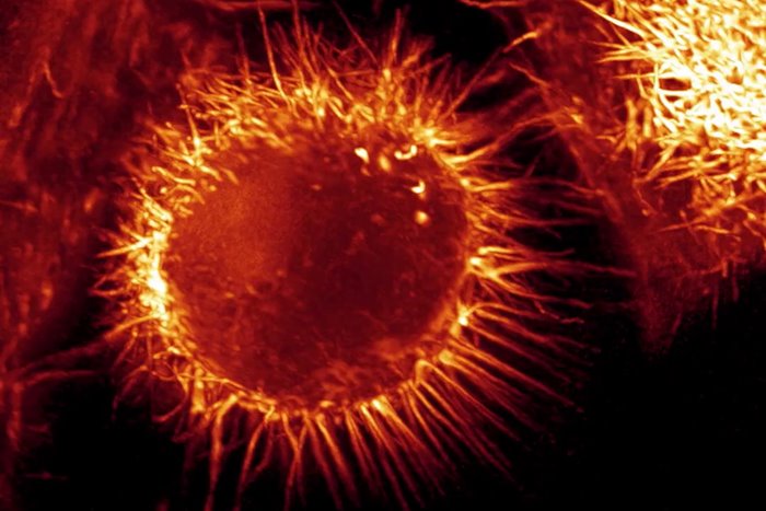
1st place: Myr John growing antlers: living coral polyps in purple and symbiotic algae in green; 10x magnification
Dr. Philippe P. Laissue from England
Second place: Tomatoes, a type of parasite, swim in the body of their host, which is a dead arthropod; 1x magnification
Dr. Richard and R. Kirby ( Dr. Richard & R. Kirby ) from England
Third place: Stylonia creates a vortex using its cilia; 10x magnification
Tommy Gunn and Jesse Gunn ( Tommy Gunn & Jesse Gunn ) from America
4th place: two freshwater tardigrades feed on another tardigrade; 10x magnification
Dr. Hunter Hines ( Dr. Hunter N. Hines ) from America
5th place: developing mouse embryo; The stage of development of the neural fold and the formation of the neural tube; 16x magnification
Dr. Kate McDole and Dr. Philip Keller ( Dr. Kate McDole & Dr. Philipp Keller ) from America
special appreciation
Iron filings in a magnetic field
Thomas Drolsum ( Thomas Drolsum ) from America
Reverse timelapse of snowflake sublimation; 4 times magnification
Caleb Foster _
A star-shaped structure called an ester consisting of mesenchyme and microtubules in an animal cell; 60 times magnification
Dr. Jesse Gatlin, Abdullah Bashar Sami, Dr. John Oakey, and Dr. April Kloxin ( Dr. Jesse Gatlin & Abdullah Bashar Sami & Dr. John Oakey & Dr. April Kloxin ) from America
hydroid; 2.5 times magnification
Raul M. Gonzalez from Mexico
discharge of the radial canal into a contractile vacuole in a single-celled animal (probably paramecium ); 40 times magnification
Edwin Lee from America
Frog developing embryo, 10th to 13th day; 4 times magnification
Dave R. Lewis from England
The waveform pattern resulting from the activity of proteins in a cell of the frog’s body, which is created spontaneously before cell division; 10x magnification
Ani Michaud, Jiaye Henry He, Dr. Bill Bement, Dr. Jan Huisken, Dr. George von Dassow ( Ani Michaud & Jiaye Henry He & Dr. Bill Bement & Dr. Jan Huisken & Dr. George von Dassow ) from America
mechanics of actin movement during cell division; 25 times magnification
Andrew Moore and Dr. Erika Holzbaur ( Andrew Moore & Dr. Erika Holzbaur ) from America
A rare type of triple cell division in myoblast culture medium (L6); 10x magnification
Dr. Patrick Charles Nahirney from Canada
formation of silver dendrites ; 3 times magnification
Wojtek Plonka from Poland
pigment cells on squid tentacles; 4 times magnification
Caroline Pritchard from America
multi-threaded worm larvae; 2 to 6 times magnification
Dr. Shinji Shimode from Japan
Neutrophils (red) inside the hindbrain of zebrafish embryos infected with a fluorescent bacterium (green); 20 times magnification
Kar Yan Soh ( Kar Yan Soh ) from New Zealand
the hatching of the smelly sen; 10x magnification
Johann Swanepoel ( Johann Swanepoel ) from South Africa
A species of ciliate swallows a string of cyanobacteria; 20 to 40 times magnification
Dr. Sally Warring ( Dr. Sally Warring ) from America