


microscopic time lapses; Nikon’s portal to the small world on the move
Nikon’s annual photomicrography competition, titled “Nikon’s Small World,” was first held in 1975 to honor the efforts of scientists who use light microscope photography in their research.
Microscopic time lapses; Nikon’s portal to the small world is on the move. In 2011 and the thirty-seventh year of holding this competition, Nikon announced that it would also accept participants’ works in a new category called “Small World in Motion.”
In the hope that this microscopic world’s astonishing details and beauty will amaze you, we look at a selection of the best works presented in the eight years of this competition.
The results presented in this section include videos and digital time-lapses recorded by various scientists with a microscope and are judged based on parameters such as the originality of the work, professional skill, visual impact, and scientific load. Videos are short and less than 25 MB, so don’t hesitate to watch them.
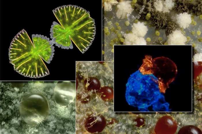
Immune system involvement with cancer cells
2014 Special Honoree: Alex Ritter, Bichang Chen, Wesley Legant, and Liang Gao from the US National Institutes of Health
Involvement of killer T lymphocyte (orange) with cancer cells (blue): T lymphocyte is a type of body immune system cell called killer T cells that directly attack virus-infected cells and cancer cells by producing a protein called perforin. They create pores in these cells and cause their death. For this reason, this type of immune response is known as the cellular immune response.
White blood cells attack the tumor.
2018 special mention: Stephen Buppart, Hahua Tu, and Eric Chaney, from the University of Illinois, USA
Development of the nervous system in zebrafish embryos
First place 2018: Elizabeth Haynes and Jay Henry, from the University of Wisconsin, USA
Sixteen hours of the development process of the sensory nervous system in zebrafish embryos: the nervous system is divided into two parts from an anatomical point of view: the central nervous system, including the brain and spinal cord, and the peripheral nervous system, including 12 pairs of brain nerve fibers and 31 pairs of spinal nerve fibers that the brain and They connect the spinal cord to other parts of the body. The peripheral nervous system consists of two main functions, sensory and motor. The sensory part directs information from the sense organs to the central nervous system, and the motor part sends nerve messages to the motor organs.
Mushroom growing hogdones
1st person 2016: The Netherlands Micropolitan Museum
Growing spores of Aspergillus niger fungus: this fungus is the cause of some infectious diseases in humans and animals. It is used in commercial applications to produce citric acid and soy sauce fermentation.
Communicating between two strands of nerve cells
2016 Special Commendation: Reynaud Renaud, from the Curie and Weizmann Institute, France
Neurons in two microcompartments bring their neurites together to communicate with each other: In the simplest terms, neurons are the primary type of nerve cells and consist of three general parts: dendrites, axons, and synaptic terminals. Branches of neurons, including dendrites and axons, are called neurites. Neurons receive nerve messages through dendrite branches and transmit them through axons. The connection point of a neuron with other neurons or with other cells is called a synapse. Any part of a neuron can synapse with any part of another neuron. The most common synapse is the synapse between the axon of one neuron and the dendrite of another neuron.
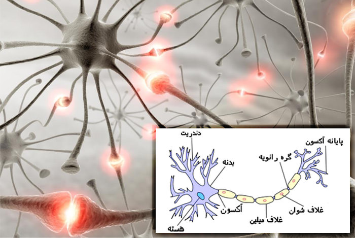
A view of the structure of a type of neuron
Cell division of a variety of green algae
Special recognition of 2016: The Netherlands Micropolitan Museum
The cardiovascular system of zebrafish
Special recognition 2015: Max Planck Institute for Molecular Genetics, Germany
The entry of malaria parasites into the mouse body by the bite of an Anopheles mosquito
2017 Special Recognition: Christine Hopp and Fotini Sinis, Johns Hopkins School of Public Health, USA
Growth of Penicillium fungi
Special recognition 2015: The Netherlands Micropolitan Museum
Heartbeat and blood vessels of a three-day-old bird embryo
First place in 2011: Anna Ferns, from the University of Oxford, England
Sweating on the skin of the finger
2nd place 2017: TIMELAPSE VISION INC, from Saitama University, Japan
The circulatory system of zebrafish parabiotic embryos
2016 Special Honoree: Elliott Hendren and Brian Lee, from Boston Children’s Hospital and Harvard Medical School, USA
Fluorescent dextran injection into the circulatory system of zebrafish parabiotic embryos to observe the typical blood flow between two organs: parabiotic is the connection of two organs or existing through surgery; in such a way that they have a standard blood circulation system.
Smartphone screen pixels running animation