

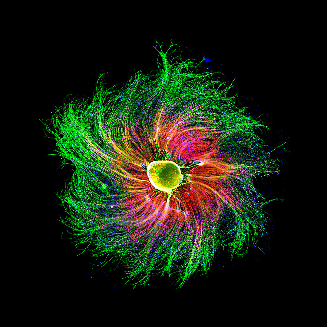
Nikon Announced The Winners Of Its 47th Small World Micrographic Photography Contest. This Year’s First Prize Was Awarded To Jason Kirk.
Nikon’s Small World competition was established in 1974 to show the beauty of photography through the eyepiece of a microscope. This event was the leading association in recognizing the art, skill, and excellence of photography in micrographic photography.
This year, this competition received about 1900 photos from 88 different countries. All images were evaluated for originality, informational content, technical skill, and visual impact.
This year’s first prize went to Jason Kirk for his striking depiction of a southern oak leaf’s veins, plant hairs, and air stomata.
Jason used various lighting techniques and design tools to capture this image, creating what the Nikon judges judged to be a masterful example of the dynamic relationship between imaging technology and artistic creativity.
First place; Jason Kirk

Using a custom microscope system that combines light transmitted through a color filter with scattered reflected light, Jason Kirk took about 200 separate images of the leaf. He stacked them to create a stunning appearance.
He used light transmitted and reflected from the sides of the leaf to highlight three vital structures in the leaf. Prominently marked in white, plant hairs are fine projections that protect the plant from adverse weather, microorganisms, and insects.
Jason has marked the air vents with purple color; These tiny pores regulate the flow of gases in the plant. The veins that carry water throughout the leaf (veins) are also visible in turquoise.
second place; Esmeralda wig
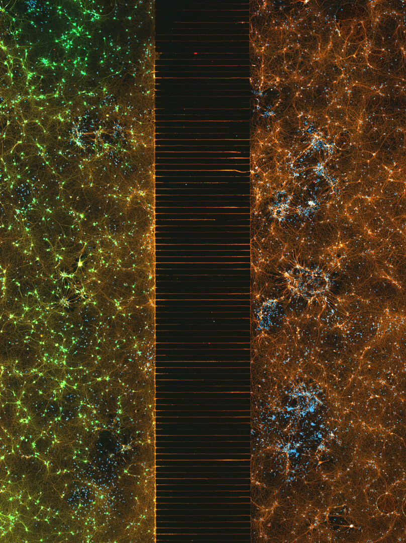

Second place went to Esmeralda Park for her image of a microfluidic device containing hundreds of thousands of network neurons. Primary nerve cells were extracted and cultured and then transfected with the virus. Esmeralda’s image shows two isolated but connected populations with different viral treatments.
third place; Frank Reiser


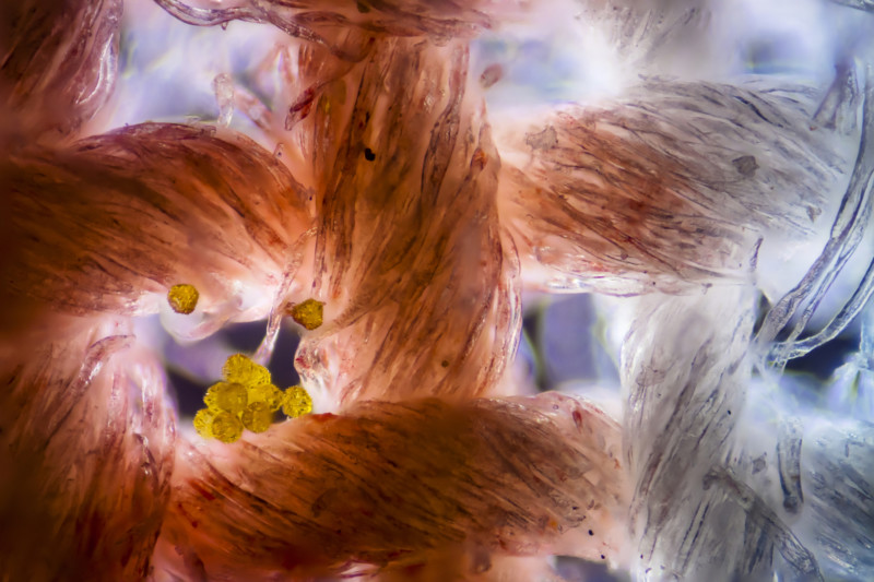
Third place went to Frank Reiser for his photograph of a hog louse’s hind legs, paws, and trachea.
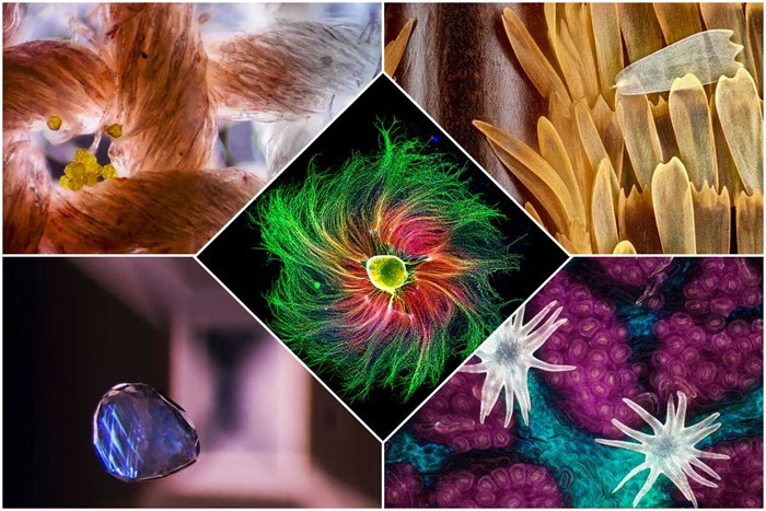
Below you can see the rest of the best pictures of this competition.

- Photographer’s name: Paula Diaz
- Subject: sensory neuron from mouse embryo

- Photographer’s name: Allison Pollock
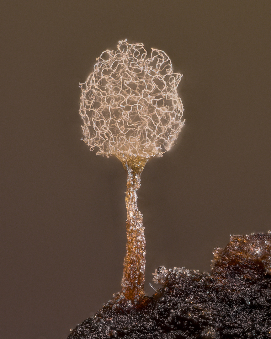
- Subject: mucous mold

- Photographer’s name: Billy Hughes
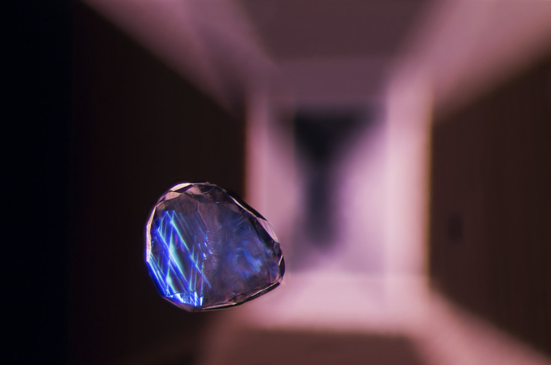
- Subject: calcite crystal suspended in Lal gemstone

- Photographer’s name: Saulius Gogis
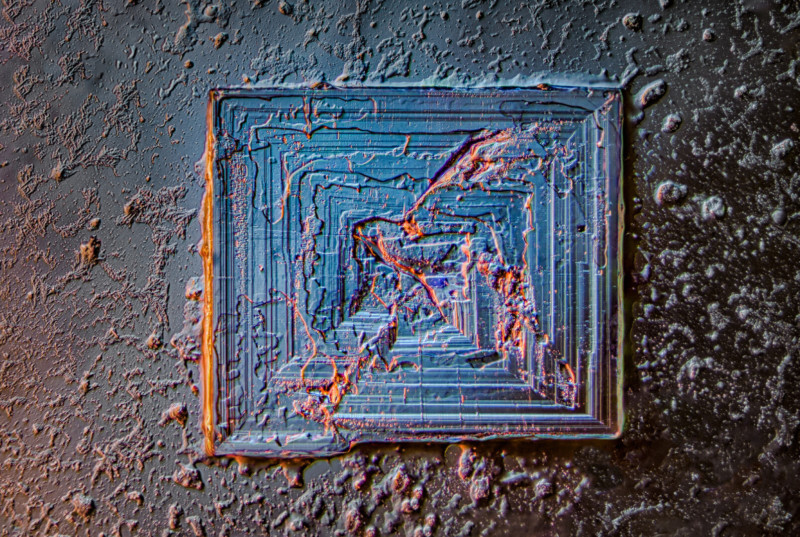
- Subject: Crystal of table salt

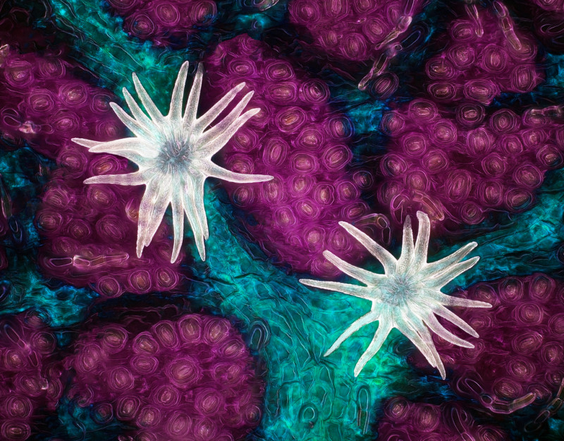
- Photographer’s name: Rohan Zhong
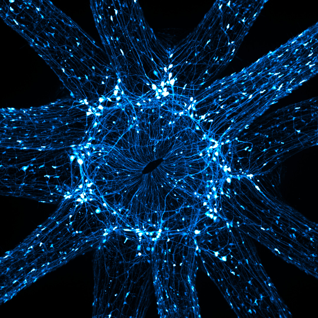
- Subject: Nerve cells around the mouth and tentacles of the starlet sea anemone
- Photographer’s name: Martin Kai Christiansen
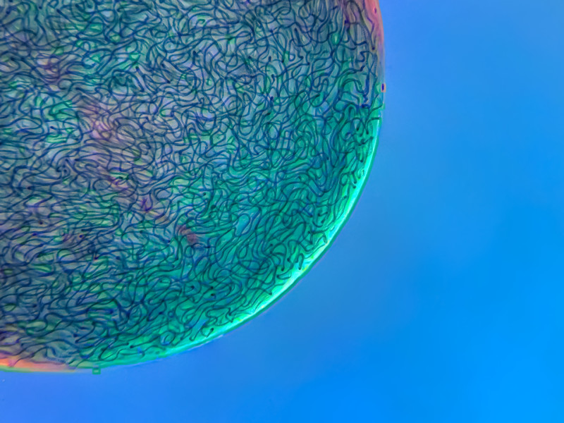
- Subject: filamentous filaments of Nostoc cyanobacteria entrapped in a gelatin matrix.

- Photographer name: Bernard Allard
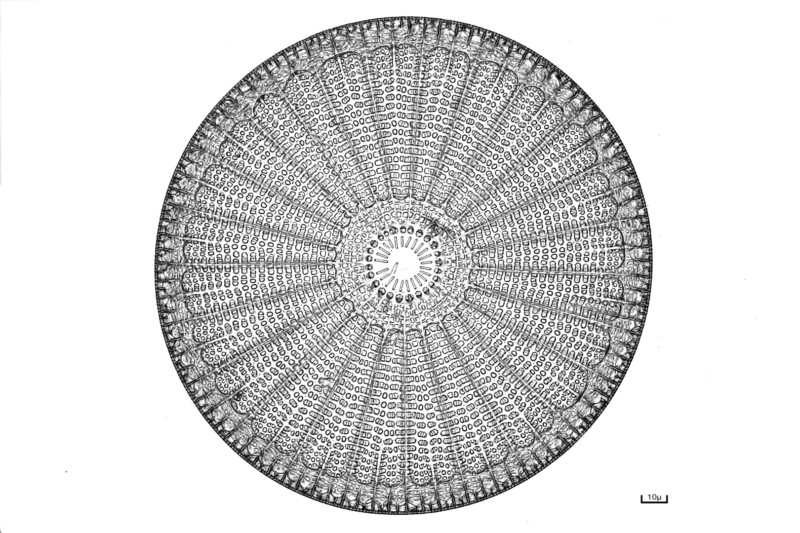
- Subject: Diatoms

- Photographer’s name: Dr. Jorn N. Hopke
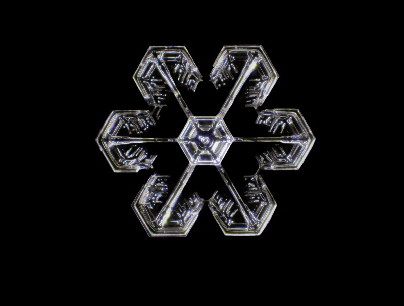
- Subject: A snowflake
- Photographer’s name: Dr. Felice Placenti
- Subject: Cotton fabric with pollen grains
- Photographer’s name: Jacob Sumbal
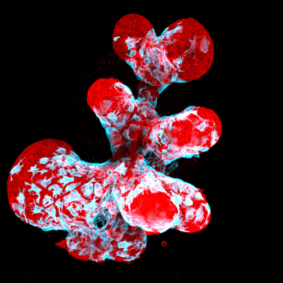
- Subject: breast organoid; The movement of myoepithelial cells (blue) on breast secretory cells (red)
- Photographer: Jason Kirk and Carlos P. Flores Suarez
- Subject: rat retinal vessels
- Photographer’s name: Sebastian Malo
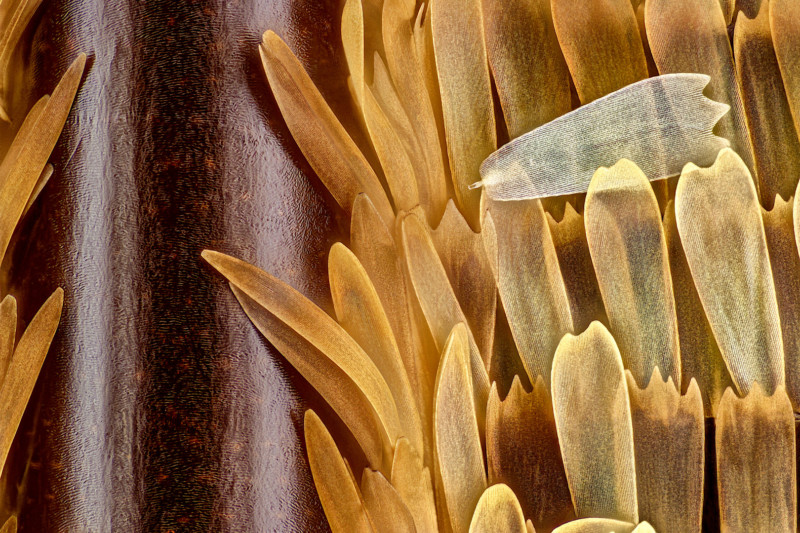
- Subject: veins and scales in the wing of a butterfly (Morpho didius)
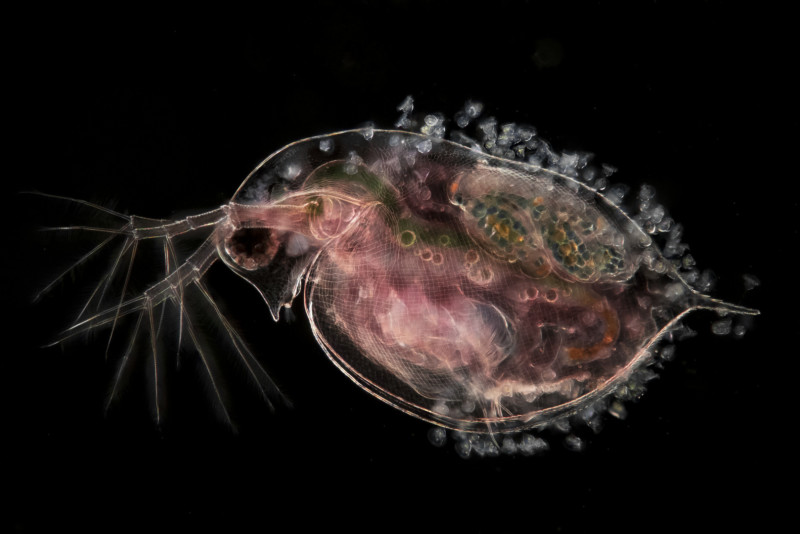

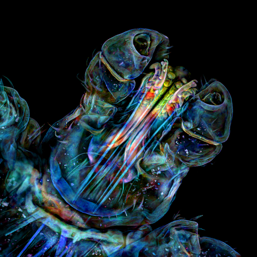
Photographer’s name: John Van Eyken
Subject: Blue Flea (Daphnia), carrying a fetus and a fairy
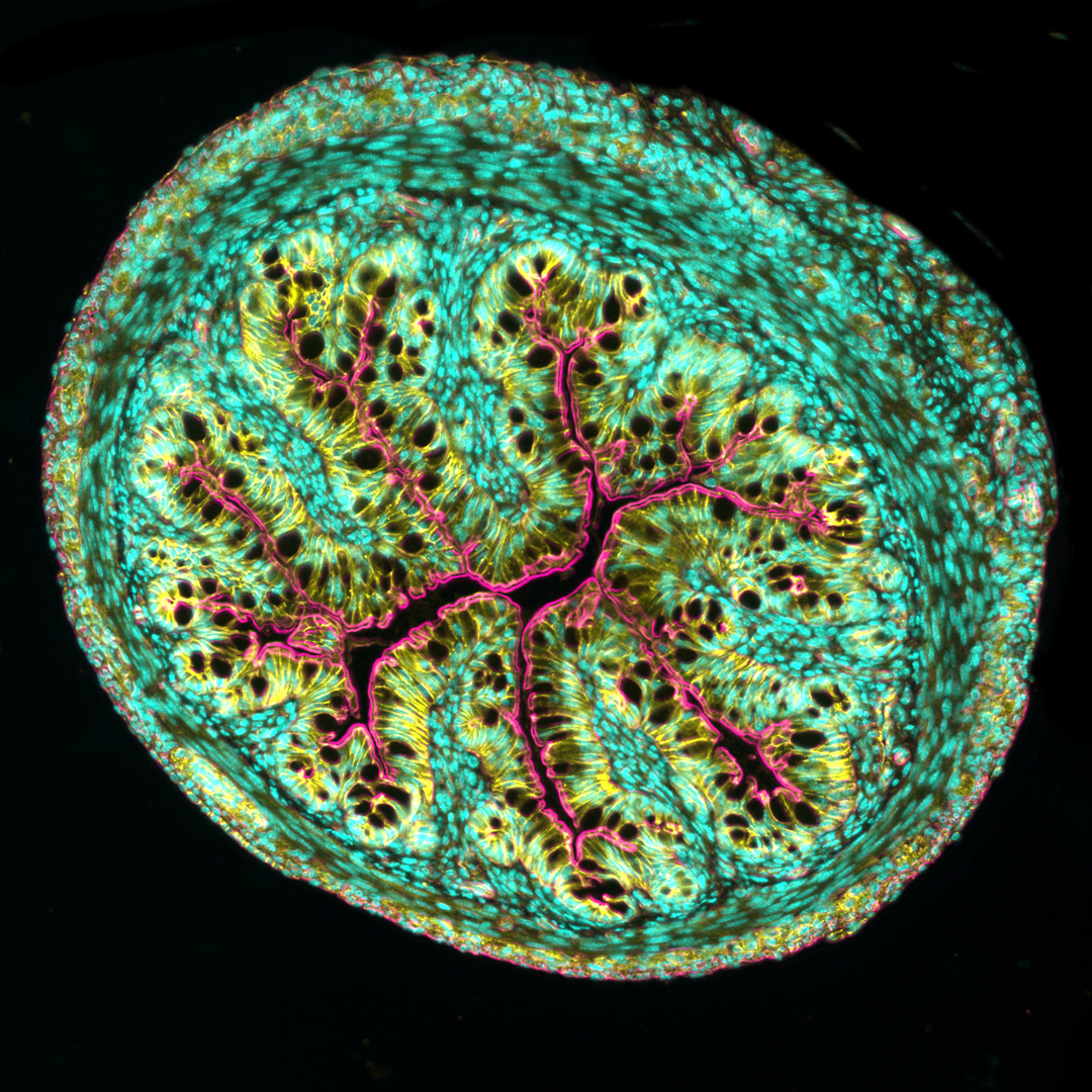
- Photographer’s name: Dr. Amy Engwick
- Subject: section of mouse intestine
- Photographer’s name: Dr. Tang Zhang and Dr. Paul Stoodley
- Subject: head
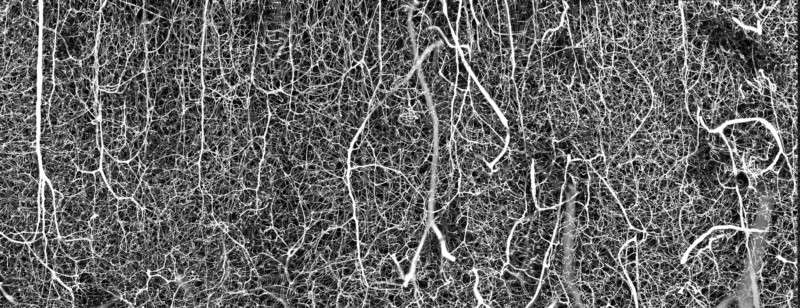

- Photographer’s name: Dr. Andrea Tedeschi
- Subject: three-dimensional vessels of the adult mouse brain (somatosensory cortex)
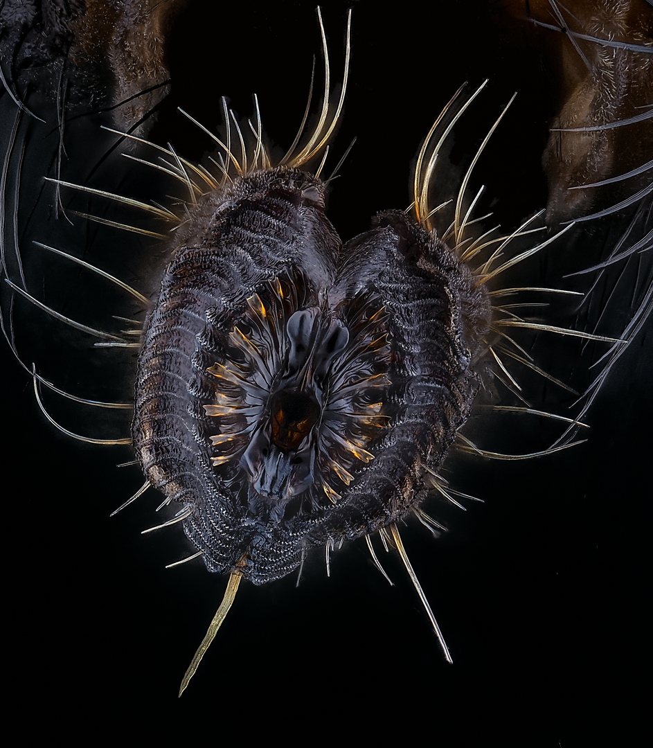

- Photographer’s name: Oliver Damm
- Subject: Proboscis of a house fly
***