

The selected works of the “Nikon Small World” competitions were introduced in the last months of 2019 in the two sections of photomicrography and microscopic time-lapses.
To watch the selected works in the microscopic time-lapse section of this competition.
“Small World in Motion” is the title of a part of “Nikon’s Small World” competition, which has hosted the works of artistic scientists every year since 2011. In this section, recorded time-lapses compete with optical microscopes. To watch these competitions’ selected works of 2019, stay with Zomit in the two areas “Selected Videos” and “Special Appreciation.”
Selected videos
1st place: Myr John growing antlers: living coral polyps in purple and symbiotic algae in green; 10x magnification
Dr. Philippe P. Laissue from England
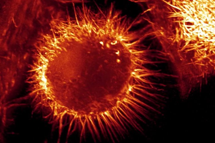
Second place: Tomatoes, a type of parasite, swim in the body of their host, which is a dead arthropod; 1x magnification
Dr. Richard and R. Kirby ( Dr. Richard & R. Kirby ) from England
Third place: Stylonia creates a vortex using its cilia; 10x magnification
Tommy Gunn and Jesse Gunn ( Tommy Gunn & Jesse Gunn ) from America
4th place: two freshwater tardigrades feed on another tardigrade; 10x magnification
Dr. Hunter Hines ( Dr. Hunter N. Hines ) from America
5th place: developing mouse embryo; The stage of development of the neural fold and the formation of the neural tube; 16x magnification
Dr. Kate McDole and Dr. Philip Keller ( Dr. Kate McDole & Dr. Philipp Keller ) from America
special appreciation
Iron filings in a magnetic field
Thomas Drolsum ( Thomas Drolsum ) from America
Reverse timelapse of snowflake sublimation; 4 times magnification
Caleb Foster _
A star-shaped structure called an ester consisting of mesenchyme and microtubules in an animal cell; 60 times magnification
Dr. Jesse Gatlin, Abdullah Bashar Sami, Dr. John Oakey, and Dr. April Kloxin ( Dr. Jesse Gatlin & Abdullah Bashar Sami & Dr. John Oakey & Dr. April Kloxin ) from America
hydroid; 2.5 times magnification
Raul M. Gonzalez from Mexico
discharge of the radial canal into a contractile vacuole in a single-celled animal (probably paramecium ); 40 times magnification
Edwin Lee from America
Frog developing embryo, 10th to 13th day; 4 times magnification
Dave R. Lewis from England
The waveform pattern resulting from the activity of proteins in a cell of the frog’s body, which is created spontaneously before cell division; 10x magnification
Ani Michaud, Jiaye Henry He, Dr. Bill Bement, Dr. Jan Huisken, Dr. George von Dassow ( Ani Michaud & Jiaye Henry He & Dr. Bill Bement & Dr. Jan Huisken & Dr. George von Dassow ) from America
mechanics of actin movement during cell division; 25 times magnification
Andrew Moore and Dr. Erika Holzbaur ( Andrew Moore & Dr. Erika Holzbaur ) from America
A rare type of triple cell division in myoblast culture medium (L6); 10x magnification
Dr. Patrick Charles Nahirney from Canada
formation of silver dendrites ; 3 times magnification
Wojtek Plonka from Poland
pigment cells on squid tentacles; 4 times magnification
Caroline Pritchard from America
multi-threaded worm larvae; 2 to 6 times magnification
Dr. Shinji Shimode from Japan
Neutrophils (red) inside the hindbrain of zebrafish embryos infected with a fluorescent bacterium (green); 20 times magnification
Kar Yan Soh ( Kar Yan Soh ) from New Zealand
the hatching of the smelly sen; 10x magnification
Johann Swanepoel ( Johann Swanepoel ) from South Africa
A species of ciliate swallows a string of cyanobacteria; 20 to 40 times magnification
Dr. Sally Warring ( Dr. Sally Warring ) from America